Advanced Electron Microscopy Core Facility
Gordon Center for Integrated Science
929 E. 57th St.
Rooms: ESB07, ESB23 (Scope room)
Chicago, IL 60637
Surgery Brain
Abbott Memorial Hall
5812 S. Ellis Ave.
Room: SB019
Chicago, IL 60637
Technical contacts:
Jotham (Joe) Austin
773-702-9091
jotham@uchicago.edu
Tera Lavoie, PhD
Technical Director
773-834-5373
tlavoie@bsd.uchicago.edu
A primary mission of the Advanced Electron Microscopy Facility is to develop techniques for preserving cellular structure with the highest degree of reliability. These techniques involve different methods for rapidly freezing our samples in order to halt structural and biochemical activity in a very short timeframe, thus preserving structure in the "live" state. Once the sample is preserved in the "live" state, it is then possible to study the ultrastructure of these samples using not only basic Electron Microscopy imaging techniques, but also state-of the-art techniques such as: 1) 3-D electron tomography, and 2) immuno-cytochemistry.
Baltec HPM01 High Pressure Freezing Machine
FEI Tecnai G2 F30 Super Twin microscope
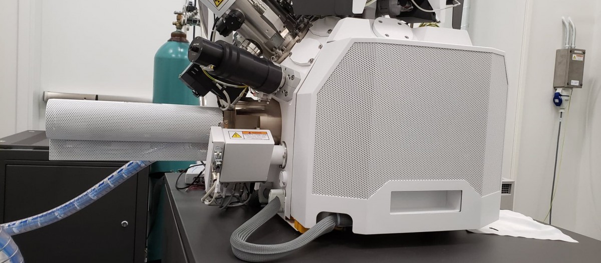
The FEI Tecnai F30 is a scanning transmission electron microscope (STEM) that forms the centerpiece of the Electron Microscopy Core Facility. The microscope can be operated at the maximum accelerating voltage of 300 kV and is capable of atomic resolution imaging in both TEM and STEM modes. The microscope uses a Schottky field emission gun as its electron source, providing the highest current density and the smallest electron probe-size, which are critical for high spatial-resolution analysis using the scanning mode.The microscope is currently equipped with a high performance CCD camera with 4k x 4k resolution for image recording, an X-ray energy dispersive spectrometer (EDX) for compositional analysis, and STEM detectors for bright-field, annular dark-field, and high-angle annual dark-field (Z-contrast) imaging.
High brightness Schottky emitter operated at 300 kV
digital CCD camera with 4k x 4k image resolution
high-resolution TEM imaging with the point-to-point resolution of 0.2 nm
STEM imaging at the spatial resolution of 0.2 nm
EDX for elemental analysis
Standard single-tilt and low-background (Be) double-tilt holders
low dose capability
tomography capability
Fischione Dual-Axis Tomography Holder Model 2040
Leica EM AFS2 Automatic Freeze-Substitution Processor
Leica UC6 Ultramicrotome with MZ6 Microscope and Accessories
Sample Equipment
Ultracut E Microtome for sectioning
Edwards Auto 306 Evaporator for carbon coating and glow discharge
Software
Gatan DigitalMicrograph
Focal-Series High-Resolution Reconstruction Package
Tomography Acquisition and Reconstruction Package
https://tomocryo.uchicago.edu/
